The International Tinnitus Journal
Official Journal of the Neurootological and Equilibriometric Society
Official Journal of the Brazil Federal District Otorhinolaryngologist Society
ISSN: 0946-5448

Google scholar citation report
Citations : 12717
The International Tinnitus Journal received 12717 citations as per google scholar report
The International Tinnitus Journal peer review process verified at publons
Indexed In
- Excerpta Medica
- Scimago
- SCOPUS
- Publons
- EMBASE
- Google Scholar
- Euro Pub
- CAS Source Index (CASSI)
- Index Medicus
- Medline
- PubMed
- UGC
- EBSCO
Volume 28, Issue 2 / August 2024
Research Article Pages:187-191
10.5935/0946-5448.20240027
Can the Facial Nerve Reduce the Loudness of Your Tinnitus?
Authors: Henk M. Koning
PDF
Abstract
Introduction: The facial nerve is a potentially target for tinnitus treatment. Objectives: The object of this study was the enduring outcome of pulsed radiofrequency of the facial nerve in patients with tinnitus and to detect predictors for a beneficial pay-off. Results: Pulsed radiofrequency of the facial nerve reduced tinnitus loudness in 25% of the patients suffering from tinnitus. Forty percent of them rated the outcome of therapy as a diminution of 50% or more. Permanent tinnitus relief after a successful pulsed radiofrequency of the facial nerve was found to be 35% at a half year follow-up. Side-effects were a 10% chance of a louder tinnitus. One patient had an epileptic attack 2 weeks after pulsed radiofrequency of the facial nerve. Patients with reduced tinnitus following pulsed radiofrequency of the facial nerve had less hearing loss at 2 and 8 kHz, normal cervical lordosis, and less disc height between the fourth and fifth cervical vertebrae compared to those with no benefit of this therapy. Conclusions: Pulsed radiofrequency of the facial nerve can be a useful alternative for patients with tinnitus. However, this beneficial effect is in most cases temporarily. Patient selection is vital for better results and less side-effects.
Keywords: Tinnitus, Facial nerve, Pulsed radiofrequency, Locus coeruleus, Nucleus paragigantocellularis.
Introduction
Cranial nerve blocks can be an auspicious intervention for patients with therapy-resistant tinnitus [1]. Neurostimulation therapies can interfere with anomalous oscillatory cortical activity and repair brain activity [2]. Pulsed Radiofrequency (PRF) is a neurostimulation technique which delivers a radiofrequency signal without producing destructive levels of heat [3]. Over the long term, PRF is an efficient and safe treatment option for cranial nerves. The Facial Nerve (FN) is a potentially target for tinnitus treatment [4]. The object of this study was the enduring outcome of PRF of the FN in patients with tinnitus and to detect predictors for a beneficial pay-off. To our knowledge is this first study of applying PRF to the FN in patients suffering from tinnitus.
Methods
Design
A looking backward study in Pain Clinic De Bilt, De Bilt, the Netherlands.
Ethical Assent
The Ethics Committee United (Nieuwegein, the Netherlands) licensed the study (W24.117, May 23, 2024).
Subjects
The study comprises every tinnitus patient who was subjected to PRF of the FN in Pain Clinic De Bilt between June 2022 and June 2024 (n = 61). No estoppel standard for PRF of the FN was used. Before therapy, a two-sided audiogram and an X-ray of the neck was obtained.
Outcome
Outcome measurements were alterations in tinnitus loudness at 7 weeks follow-up and period of enduring tinnitus repression after PRF of the FN.
Adverse Effects
Side effects were enrolled immediately after RRF of the FN and at 7 weeks follow-up.
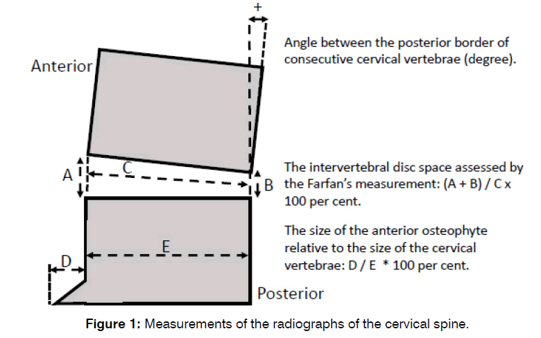
X-ray of the neck
Elucidate the measurements of the X-ray of the neck (Figure 1).

Figure 1: Measurements of the radiographs of the cervical spine.
PRF of the FN
The nerve block was carried out by an anesthesiologist. The patient is put in a supine position with the head turned away from the side to be blocked. The mastoid process is then identified by palpation. After decontamination, a 22-gauge, 60 mm-long needle with a 5 mm active tip was put in the middle of the posterior boundary of the mandibular ramus and the frontier border of the mastoid process, just atop of the lowest end of the earlobe. Then, the needle was preceded for 20-25 mm to the expected stylomastoid foramen. After aspiration for blood, PRF at 42 V, 2 Hz, and 10 milliseconds for 10 minutes was administered. Following the procedure, patients were kept under observation for 30 minutes. The result of the treatment was assessed 7 weeks post treatment.
Data Assessment
Data included tinnitus characteristics (duration of tinnitus, presence of hearing loss, vertigo or balance disorders), benefit at 7 weeks follow-up on a 4-point scale (none [0%], slight [< 25%], moderate [25% -50%], good [50% or greater]), and the period of the benefit. Seven weeks following the procedure, other therapy was proposed. Patients with a beneficial effect and no recurrence nor other therapy, were invited for an interview to assess the duration of benefit. In August 2024, a survey by a nonparticipant was accomplished.
Statistical Analysis
Analysis was carried out with Minitab 18 (Minitab Inc., State College, PA, USA) using Student’s t-test, χ2 test, survival analysis, stepwise regression and discriminant analysis. A P-value fewer than 0.05 was statistically meaningful.
Results
In a 2-year period, 61 tinnitus patients were treated with PRF of the FN (Table 1). introduces the clinical characteristics. Sixteen (25%) reported benefit at the 7-week follow-up. The patients valued their benefit as 40% good, 53% moderate, and 7% slight. The loudness of tinnitus magnified in 10% of the treated patients. One patient had an epileptic attack 2 weeks after therapy. The follow-up of successful treated patients was up to 23 months postoperative. Permanent tinnitus relief after a successful PRF of the FN was found to be 35% at a half year follow-up (Figure 2).
| Prevalence | Median | Q1 – Q3 | |
|---|---|---|---|
| Age (year) | 54 | 46 – 64 | |
| Gender (male) | 61% | ||
| Unilateral tinnitus | 31% | ||
| Self-perceived hearing loss | 62% | ||
| Cervical pain | 75% | ||
| Period of tinnitus (year) | 6 | 1 – 15 | |
| Hearing loss (dB) at: | |||
| 250 Hz | 15 | 7 – 25 | |
| 500 Hz | 15 | 5 – 25 | |
| 1 kHz | 10 | 8 – 30 | |
| 2 kHz | 20 | 10 – 34 | |
| 4 kHz | 30 | 19 – 50 | |
| 8 kHz | 43 | 20 – 60 |
Table 1: Clinical Characteristics of the patients with tinnitus.

Figure 2: Kaplan-Meier graph to show indicating the odds of permanent tinnitus relief in successfully treated patients (n=11) after PRF of the facial nerve. Two patients did not respond to our invitation for a question-and-answer session to value time of improvement.
Patients with a successful response to therapy were compared to those who had not (Table 2). Hearing loss at 2 and 8 kHz, cervical lordosis, and disc height between the fourth and fifth cervical vertebrae were statistically significant different between both groups. Patients with reduced tinnitus following PRF of the FN had less hearing loss at 2 and 8 kHz, normal cervical lordosis, and less disc height between the fourth and fifth cervical vertebrae compared to those with no benefit of this therapy.
| Positive effect of therapy (n=15) | No effect of therapy (n=46) | P-value | |||||
|---|---|---|---|---|---|---|---|
| Prev. | Mean | SEM | Prev. | Mean | SEM | ||
| Age (year) | 52 | 3.8 | 55 | 1.9 | 0.57 | ||
| Gender (male) | 53% | 63% | 0.504 | ||||
| Unilateral tinnitus | 47% | 26% | 0.135 | ||||
| Self-perceived hearing loss | 47% | 67% | 0.15 | ||||
| Cervical pain | 67% | 78% | 0.365 | ||||
| Age at the start of tinnitus (year) | 47 | 4.2 | 45 | 3.4 | 0.735 | ||
| Hearing loss (dB) at: | |||||||
| 250 Hz | 18 | 3.6 | 19 | 2.8 | 0.844 | ||
| 500 Hz | 17 | 4 | 19 | 3 | 0.708 | ||
| 1 KHz | 16 | 3.3 | 21 | 3 | 0.218 | ||
| 2 KHz | 16 | 3.5 | 26 | 3.2 | 0.045 Sign. | ||
| 4 KHz | 28 | 4.4 | 38 | 3.6 | 0.092 | ||
| 8 KHz | 31 | 6.2 | 47 | 4.2 | 0.048 Sign. | ||
| Angle between vertebrae C2 and C6 (degrees): | 10 | 1.8 | 5 | 1.6 | 0.041 Sign. | ||
| Farfan’s measurement of disc space height (%): | |||||||
| C2-C3 | 42 | 1.6 | 39 | 1.5 | 0.149 | ||
| C3-C4 | 40 | 1.9 | 35 | 1.5 | 0.065 | ||
| C4-C5 | 39 | 1.5 | 33 | 1.4 | 0.008 Sign. | ||
| C5-C6 | 30 | 2 | 27 | 1.4 | 0.265 | ||
| C6-C7 | 27 | 2.6 | 26 | 1.4 | 0.673 | ||
| Size of anterior osteophyte (%) at: | |||||||
| C3 | 8 | 1.4 | 9 | 0.9 | 0.36 | ||
| C4 | 12 | 1.1 | 13 | 1.1 | 0.529 | ||
| C5 | 16 | 1.3 | 19 | 1.3 | 0.076 | ||
| C6 | 16 | 1.9 | 15 | 1.1 | 0.572 | ||
Table 2: Patients with a positive effect of therapy of the facial nerve on their tinnitus at 7 weeks were compared with non-responders.
The patient group with a better result of PRF of the FN was identified using discriminant analysis. Patients with a Farfan measurement of the disc between the 4th and 5th cervical vertebrae above 36% had 34% chance on a positive effect compared to 16% in patients not fulfilling these criteria. Also, there was a difference in side-effects between these groups of patients, 7% compared to 16%.
Discussion
PRF of the FN reduced tinnitus loudness in 25% of the patients suffering from tinnitus. Forty percent of them rated the outcome of therapy as a diminution of 50% or more. Permanent tinnitus relief after a successful PRF of the FN was found to be 35% at a half year follow-up. Side-effects were a 10% chance of a louder tinnitus. One patient had an epileptic attack 2 weeks after PRF of the FN. Patients with reduced tinnitus following PRF of the FN had less hearing loss at 2 and 8 kHz, normal cervical lordosis, and less disc height between the fourth and fifth cervical vertebrae compared to those with no benefit of this therapy.
The FN is potentially target for tinnitus treatment. Nerve block of the FN has reported to be beneficial in reducing tinnitus intensity [4]. Also, we conclude that PRF of the FN can diminish tinnitus complaint is a restricted part of the tinnitus population. However, this beneficial effect is in most cases temporarily. The chance of a better result and less side-effects can be improved by selecting patients with a Farfan measurement of the disc between the 4th and 5th cervical vertebrae above 36%.
The mechanism of action of PRF on the FN could be an effect on the nerve itself or a central effect by interruption of anomalous oscillatory cortical activity [2]. A damaged FN is associated with hearing loss, tinnitus, imbalance, and hypersensitivity to sound [5]. In our study, tinnitus patients with an improvement by PRF of the FN had less hearing loss at 2 and 8 kHz compared to those with no benefit of this therapy. Therefore, a direct action of PRF on the FN itself we do not consider likely and a central effect is more feasible.
The FN has 3 distinct brain stem nuclei (motor nucleus of FN, Nucleus of Solitary Tract (NTS), and superior salivatory nuclei for motor, taste, and salivation or lacrimation, respectively) situated in the pons. The second infratemporal branch of the FN is the nerve to stapedius muscle, responsible for dampening vibrations and protecting the hearing apparatus when exposed to loud sounds. Dysfunctional stapedius contraction can directly stimulate the cochlea and causes symptoms of tinnitus [6,7]. It is possible that activation of the motor nucleus of the FN by PRF restore the dysfunctional movement of the stapedius muscle.
Sensory information via afferent fibers of the FN enters the brainstem and forms the solitary tract and then synapse with NTS [8]. The NTS coordinates autonomic information to the nucleus Paragigantocellularis (PGi) and to the central nucleus of the amygdala (CeA) [9]. The major input to the Locus Coeruleus (LC) comes from the Pgi (excitatory) and the nucleus prepositus hypoglossi (inhibitory) [10]. This gives PRF of the FN a direct excitatory pathway to the LC. The LC is the Noradrenergic (NE) modulator of the central nervous system, responsible for adapting cortical circuits to task demands [11]. LC-NE activation facilitates the representation of sensory signals by inhibiting spontaneous activity more than sensory-evoked responses, thus effectively enhancing the signal-to-noise ratio [12]. Noradrenergic modulation gives a specific disinhibitory signal to the auditory system which can reduce the loudness of tinnitus [13].
Our study concludes that PRF of the FN can reduce the loudness of tinnitus only in a small part of the patients (25%). In these tinnitus patients, PRF of the FN activates the excitatory pathway from the NST to the LC leading to an inhibitory signal to the auditory system. Hearing loss at 2 and 8 kHz, less cervical lordosis, and disc degeneration between the fourth and fifth cervical vertebrae can impede the effect of PRF of the FN. It could well be that the mechanism responsible for this interaction is situated in the nucleus paragigantocellularis as much of the somatosensory and mechanoreceptor input, auditory input via the inferior colliculus, sympathetic afferent input, and input from the FN comes here together.
This study has limitations. First, the retrospective study-design can limit the certainty of the conclusions. The second restriction is the total of patients involved in this study. A prospective study with a larger number of patients is recommended.
Conclusion
PRF of the FN can be a useful alternative for patients with tinnitus. However, this beneficial effect is in most cases temporarily. Patient selection is vital for better results and less side-effects. A prospective study should further evaluate these findings.
Conflicts of Interest and Source of Funding
The authors declare no conflict of interest.
References
- Koning HM, van Hemert FJ. Pulsed Radiofrequency of the Vagal Nerve for Tinnitus-A Case-Study. Int Tinnitus J. 2021 Dec 28;25(2):172-5..
- Adair D, Truong D, Esmaeilpour Z, Gebodh N, Borges H, Ho L, et al. Electrical stimulation of cranial nerves in cognition and disease. Brain Stimul. 2020;13(3):717-50.
- Yadav YR, Nishtha Y, Sonjjay P, Vijay P, Shailendra R, Yatin K. Trigeminal Neuralgia. Asian J Neurosurg. 2017;12(4):585-597.
- Sirh SJ, Sirh SW, Mun HY, Sirh HM. Integrative treatment for tinnitus combining repeated facial and auriculotemporal nerve blocks with stimulation of auditory and non-auditory nerves. Front.Neurosci. 2022 Feb 28;16:758575.
- Bartindale M, Heiferman J, Joyce C, Balasubramanian N, Anderson D, Leonetti J. The natural history of facial schwannomas: a meta-analysis of case series. J Neurol Surg B Skull Base. 2019;80(05):458-68.
- Onishi ET, Coelho CC, Oiticica J, Figueiredo RR, Guimarães RD, Sanchez TG, et al. Tinnitus and sound intolerance: evidence and experience of a Brazilian group. Braz J Otorhinolaryngol. 2018;84:135-49.
- Shokri T, Azizzadeh B, Ducic Y. Modern management of facial nerve disorders. Semin Plast Surg. 2020;34(4):277-285.
- Møller AR. Sensorineural tinnitus: its pathology and probable therapies. Int J Otolaryngol. 2016;2016:2830157.
- Reyes BA, Van Bockstaele EJ. Divergent projections of catecholaminergic neurons in the nucleus of the solitary tract to limbic forebrain and medullary autonomic brain regions. Brain Res. 2006;1117(1):69-79.
- Cakmak YO, Apaydin H, Kiziltan G, Gündüz A, Ozsoy B, Olcer S, et al. Rapid alleviation of Parkinson’s disease symptoms via electrostimulation of intrinsic auricular muscle zones. Front Hum Neurosci. 2017;11:338.
- De Cicco V, Tramonti Fantozzi MP, Cataldo E, Barresi M, Bruschini L, Faraguna U, et al. Trigeminal, visceral and vestibular inputs may improve cognitive functions by acting through the locus coeruleus and the ascending reticular activating system: a new hypothesis. Front Neuroanat. 2018;11:130.
- Jacobs HI, Priovoulos N, Poser BA, Pagen LH, Ivanov D, Verhey FR, et al. Dynamic behavior of the locus coeruleus during arousal-related memory processing in a multi-modal 7T fMRI paradigm. Elife. 2020;9:e52059.
- McBurney-Lin J, Lu J, Zuo Y, Yang H. Locus coeruleus-norepinephrine modulation of sensory processing and perception: A focused review. Neurosci Biobehav Rev. 2019;105:190-199.
Department of Pain therapy, Pain Clinic De Bilt, De Bilt, The Netherlands
Send correspondence to:
H.M. Koning
Department of Pain therapy, Pain Clinic De Bilt, De Bilt, The Netherlands, E-mail: hmkoning@pijnkliniekdebilt.nl
Tel: 0031302040753
Paper submitted on Aug 16, 2024; and Accepted on Aug 21, 2024
Citation: Henk M. Koning. Can the Facial Nerve Reduce the Loudness of Your Tinnitus?. Int Tinnitus J. 2024;28(1):187-191


