The International Tinnitus Journal
Official Journal of the Neurootological and Equilibriometric Society
Official Journal of the Brazil Federal District Otorhinolaryngologist Society
ISSN: 0946-5448

Google scholar citation report
Citations : 12717
The International Tinnitus Journal received 12717 citations as per google scholar report
The International Tinnitus Journal peer review process verified at publons
Indexed In
- Excerpta Medica
- Scimago
- SCOPUS
- Publons
- EMBASE
- Google Scholar
- Euro Pub
- CAS Source Index (CASSI)
- Index Medicus
- Medline
- PubMed
- UGC
- EBSCO
Volume 27, Issue 2 / December 2023
Research Article Pages:238-241
10.5935/0946-5448.20230036
Surgical option for External Auditory Canal Cholesteatoma: A case report
Authors: Fathimath Ruhusha Jameel*, Asma Abdullah, Noor Dina Hashim, Zara Nasseri, Wan Nabila Wan Mansor
PDF
Abstract
External Auditory Canal Cholesteatomas (EACC), is an exceptionally rare condition with a prevalence of only 0.1-0.5% among new patients1. EACC are known to possess bone eroding properties, causing a variety of complications, similar to the better-known attic cholesteatomas. We describe here the novel surgical management of a case of EACC. She is 38-year-old female who presented with otorrhea for 6 months. Clinical examination and radiological investigations suggested the diagnosis of an external auditory canal cholesteatoma. The patient underwent modified radical mastoidectomy with type 1 tympanoplasty with meatoplasty. Post-operatively, the patient showed marked clinical improvement.
Keywords: External auditory canal cholesteatoma, Keratosis obturans, modified radical mastoidectomy.
Introduction
External auditory canal cholesteatoma is a rare otological disease with an incidence of 1:1000 of all new patients in otologic practice [1]. It is quite common to misdiagnose EACC as keratosis obturans or otomycosis. Symptoms and signs of focal erosion, inflammation and keratin retention are not pathognomonic for EACC [2] An EACC can erode bone, causing complications such as the invasion of the mastoid cavity, facial nerve paralysis, labyrinthine fistulae, erosion of the temporomandibular joint, invasion of the skull base, meningitis, and intracranial abscesses, not unlike an attic cholesteatoma.
Case Report
A 38 female lady presented to ENT clinic with complaints of right sided otalgia and otorrhea for the past 6 months. The otalgia was mild, dull in nature and did not affect her daily activities. Ear discharge was scanty in amount and with a putrid smell. She did not give any history of loss of hearing, aural fullness, vertigo, nausea or vomiting. There was no history of previous ear infections.
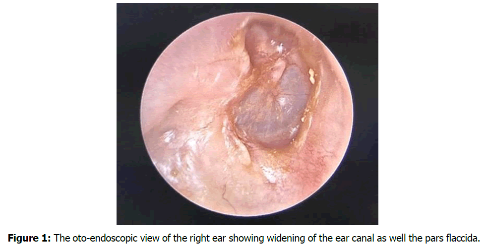
On otoscopic examination the external auditory canal was widened with thickened and dull tympanic membrane. Pars flaccida was widened as well with TOS grade IV retraction. No keratin or granulation tissue was seen. There was bony erosion of the inferior part of the EAC. Facial nerve was intact. Tuning fork examination and pure tone audiometry showed right sided mild conductive hearing loss. The rest of the otolaryngologist, general and systemic and examination were unremarkable Figure 1.

Figure 1: The oto-endoscopic view of the right ear showing widening of the ear canal as well the pars flaccida.
A High-Resolution Computed Tomography (HRCT) of the temporal bone was taken which showed soft tissue thickening within the right middle ear canal with part of the malleus covered by the soft tissue. Epitympanic recess was also partially filled with the mucosal thickening. The floor of the mastoid lateral to the tympanic membrane was eroded. Right mastoid air cells were also fully filled with soft tissue lesion Figure 2.

Figure 2: High resolution CT scan showing right mastoid air cells filled with soft tissue and erosion of the posterior wall of the external auditory canal (white arrow).
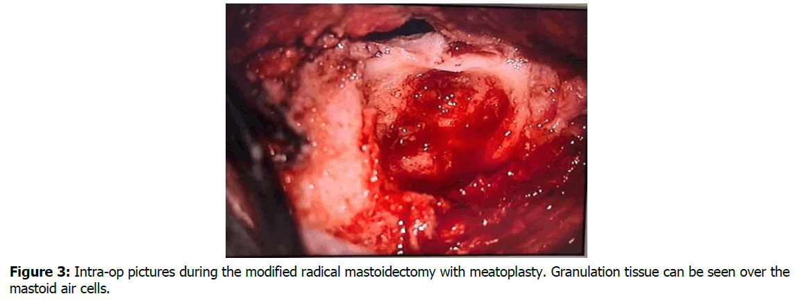
The patient underwent modified radical mastoidectomy with type 1 tympanoplasty with meatoplasty under general anesthesia. Intra-operatively post-auricular incision was given 1 cm posterior to the posterior sulcus and posteriorly based flap raised. This was followed by cortical mastoidectomy, superiorly till the superior temporal line, inferiorly till mastoid tip, anteriorly till posterior wall of the EAC and posteriorly till sigmoid sinus. Granulation tissue was seen over the mastoid air cells. Body and short process of incus, head of malleus and suspensory ligament were identified and preserved. Granulation tissue was seen over the epitympanum and over the ossicles. Facial nerve was identified and preserved anatomically and physiologically. Posterior wall of the EAC was brought down and meatoplasty performed. Temporalis fascia was harvested and placed by on-lay method. Mastoid cavity packed with bismuth and iodoform paraffin paste. Extending to the external auditory canal. Wound was closed in layers and mastoid dressing applied.
The post-operative period was uneventful and patient was discharged home on the second post-op day.
Discussion
The first reported case of EACC was made by Tonybee [3] in 1850 in which he described a lesion of epidermoid scales arising from the external auditory meatus and eroding into the mastoid. However, when the disease is limited to the ear canal there is a need to differentiate EACC from keratosis obturans as both disease produces accumulation of keratin which was highlighted by Pierpergerdes et al [4]. Previously the formation of EACC was via “keratinization in situ” which is a reduction in migratory capacity of epithelium which from the malleus towards the epithelium [5] Figure 3. However there is recent experimental data by Bonding P et al [6] which falsified this traditional pathogenetic model.

Figure 3: Intra-op pictures during the modified radical mastoidectomy with meatoplasty. Granulation tissue can be seen over the mastoid air cells.
The cardinal symptoms of EACC are unilateral otorrhea, with few reports of unilateral otalgia as well [7]. These findings distinguish EACC from keratosis obturans, which in turn, typically occurs bilaterally with intensive otalgia combined with hyperacusis which is not a typical complaint for EACC [7]. The first classification for staging of EACC was proposed by Naim et al [2] which is based on pathological, clinical and radiological evidence. In the earlier stages (stages I and II) there is no destruction of the bony canal, whereas bony destruction of EAC and involvement of adjacent structures are seen in advanced stages (stages III and IV).
The primary objective in managing EACC is similar to that of middle ear cholesteatoma. It is crucial to completely eradicate the cholesteatoma and preserve normal structures as much as possible to restore epithelial migration in the EAC. In the earlier stages, when EACC does not involve bony destruction, conservative treatment is recommended. If these measures are proven to be insufficient to control the clinical symptoms, or if patients are in the advanced stage with bony canal destruction or damage to adjacent anatomical structures, surgery should be considered. The surgical goal is to excise affected areas with a clear margin of healthy epithelium, carefully removing eroded skin and damaged bone [8]. When the mastoid air cells are invaded, a modified radical mastoidectomy is indicated with preservation of the tympanic membrane and ossicles Table 1. The following Table 2 summarizes the proposed staged treatment options for EACC in relation to the degree of invasion and extension in the tympanomastoid cell system as proposed by Patrick et al in their meta-analysis of external auditory canal cholesteatoma.
| Stage 1 | Stage II | Stage III | Stage IV |
|---|---|---|---|
| Hyperplasia and hyperemia of the auditory meatal epithelium | No destruction of the bony canal. Accumulation of keratin debris | Destruction of the bony canal with sub sequestrated bone | Destruction of the adjacent anatomical structures |
| a. Intact epithelial surface b. Excavation of the defective epithelium with apparent bony canal |
Subclass M: Mastoid Subclass S: Skull base with sigmoid sinus Subclass J: Temporal-mandibular joint Subclass F: Facial nerve |
Table1: The classification of EACC as proposed by Naim et al.
| Stage I: hyperkeratosis | Stage II: Periosteitis | Stage III: Bone invasion | Stage IV: Destruction of neighboring structures |
|---|---|---|---|
| Topical antibiotic and anti-inflammatory ointments | Topical therapy and eventually biopsy (stage IIa), full curettage of the lesion or meato-canaloplasty if superficial bone erosion is present (stage IIb) | Meato-canaloplasty (mostly under general anesthesia) | Extended otomastoid surgery or temporomandibular joint revision |
Table 2: Treatment options for EACC as proposed by Patrick et al.
Conclusion
External auditory canal cholesteatoma is a rare disease entity which is not very well understood. It is difficult to diagnose the disease in the earlier stages and usually patients present with advanced disease requiring surgical intervention. Pre-operative radiological evaluation and surgical exploration leads to a correct and practical staging of external auditory canal cholesteatoma. The treatment is by surgery with reconstruction as per the stage and degree of invasion of external auditory canal cholesteatoma.
References
- Hartley C, Birzgalis AR, Lyons TJ, Hartley RH, Farrington WT. External ear canal cholesteatoma: case report. Ann Otol Rhinol Laryngol.1995;104(11):868-70.
- Anthony PF, Anthony WP. Surgical treatment of external auditory canal cholesteatoma. Laryngoscope. 1982;92(1):70-5.
- Naim R, Linthicum Jr F, Shen T, Bran G, Hormann K. Classification of the external auditory canal cholesteatoma. Laryngoscope. 2005;115(3):455-60.
- Toynbee J. Specimens of molluscum contagiosum developed in the external auditory meatus. Lond Med Gaz. 1850;46(11):261-4.
- Piepergerdes JC, Kramer BM, Behnke EE. Keratosis obturans and external auditory canal cholesteatoma. Laryngoscope. 1980;90(3):383-91.
- Makino K, Amatsu M. Epithelial migration on the tympanic membrane and external canal. Archives of oto-rhino-laryngology. 1986 Mar;243:39-42.
- Bonding P, Ravn T. Primary cholesteatoma of the external auditory canal: is the epithelial migration defective?. Otol Neurotol. 2008;29(3):334-8.
- Dubach P, Mantokoudis G, Caversaccio M. Ear canal cholesteatoma: meta-analysis of clinical characteristics with update on classification, staging and treatment. Curr Opin Otolaryngol Head Neck Surg. 2010;18(5):369-76.
1Department of Otorhinolaryngology Head and Neck Surgery, Faculty of Medicine, Universiti Kebangsaan Malaysia, Kuala Lumpur, Malaysia
2Hospital Canselor Tuanku Muhriz, Jalan Yaacob Lathif, Bandar Tun Razak, Kuala Lumpur
3Centre of Hearing and Speech ( Pusat-HEARS), Universiti Kebangsaan Malaysia, Kuala Lumpur, Malaysia
Send correspondence to:
Fathimath Ruhusha Jameel
Department of Otorhinolaryngology, Universiti Kebangsaan Malaysia Medical Centre, Jalan Yaacob Latif, Bandar Tun Razak, 56000 Kuala Lumpur, Malaysia
E-mail: ruhshajameel29@gmail.com
Paper submitted on November 13, 2023; and Accepted on December 04, 2023
Citation: Jameel RF. Surgical Option for External Auditory Canal Cholesteatoma: A Case Report. Int Tinnitus J. 2023;27(2):238-241.


